The task of understanding the inner workings of the brain has fascinated both philosophers and scientists for centuries. Aristotle proposed that the brain is where spirit resides. Leonardo da Vinci drew anatomical depictions of the brain with wax embedding. And Santiago Ramón y Cajal, with his 1906 Nobel Prize-winning work on the cellular structure of the nervous system, made one of the first breakthroughs that led to modern neuroscience as we know it.
Using a new way to visualize individual cells called Golgi staining, a method pioneered by Nobel co-winner Camillo Golgi, and microscopic examination of brain tissue, Cajal established the seminal neuron doctrine. This principle states that neurons, among the main types of brain cells, communicate with one another via the gaps between them called synapses. These findings launched a race to understand the cellular composition of the brain and how brain cells are connected to one another.
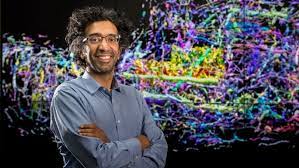
Neuroscience has since experienced a rapid explosion of new experimental tools. Jumping forward 100 years to today, modern tools called neurotechniques, which include brain mapping, have given neuroscientists a way to closely inspect every component of the brain. My lab has been utilizing these brain mapping tools to understand what cell types make up the brain and how they contribute to the creation of cognition.
Scientists first need to label, or visualize, a specific cell type. The process is like finding a needle in a haystack – it would be a lot easier to find if the needle, or cell type, glowed. This can be done with either genetic or immunostaining methods. The genetic method takes advantage of animals, like mice, that can be genetically engineered so only the target cell type is visible under specific fluorescent lights. Immunostaining methods, on the other hand, render brain samples transparent with a special chemical treatment and use antibodies to label the target cell type with a fluorescent tag.
The next step is to image the whole brain using microscopy techniques that allow scientists to view parts too small for the naked eye to see. Specialized microscopy tools can take snapshots, or tiles, of the entire brain. Stitching these image tiles together can reconstruct an intact 3D volume like a photo mosaic. It’s like building a Google map of the brain: By combining millions of individual street photos, you can zoom in to see each street corner and zoom out to see an entire city.
Unsurprisingly, this type of 3D imaging creates very large datasets. Even though a mouse brain is smaller than a human fingertip, the size of these datasets can easily reach between a few hundred gigabytes to a terabyte. Luckily, remarkable advances in computer equipment and software have made large-scale data analysis possible. Artificial intelligence algorithms in particular have enabled scientists to detect many different cell features in the brain, such as cell shape and size, as well as the processes they undergo.
Source : https://www.popsci.com/technology/human-brain-map-technology/